Home
/ Plant Cell Transmission Electron Microscope Images / Tem Of Plant Cell - Transmission electron microscopy <ul><li>in a conventional transmission electron microscope, a thin specimen is irradiated with an electron.
Plant Cell Transmission Electron Microscope Images / Tem Of Plant Cell - Transmission electron microscopy <ul><li>in a conventional transmission electron microscope, a thin specimen is irradiated with an electron.
Plant Cell Transmission Electron Microscope Images / Tem Of Plant Cell - Transmission electron microscopy <ul><li>in a conventional transmission electron microscope, a thin specimen is irradiated with an electron.. Thus, the atmospheric transmission is particularly important for the spread of epiphytic bacteria. A very high resolution for observing and analyzing the composition of elementary structures. What is transmission electron microscopy ? Difference between electron beam and light. They have a much higher resolution (can provide clearer and more detailed images).
Transmission electron microscopy <ul><li>in a conventional transmission electron microscope, a thin specimen is irradiated with an electron. The final image produced from an electron microscope is always when you look at animal or plant cells under the electron microscope, you can see a lot more detail. At a maximum potential magnification of 1 nanometer, tems are the most powerful microscopes. As its name suggests, electron microscopy is not based on photons but on electrons. Transmission electron microscopy and electron diffraction.
Tem Plant Cell High Resolution Stock Photography And Images Alamy from c8.alamy.com Scanning electron microscope and transmission electron microscope. For imaging electrons scatterring ,heavy metals like uranium and lead are used and thus give contrast. Swagelok center for surface analysis of materials (scsam) case western reserve university. They have a much higher resolution (can provide clearer and more detailed images). What is transmission electron microscopy ? See more ideas about electron microscope, microscopic photography, microscopic. Electron microscopes have a much higher resolution than light microscopes. Colorized transmission electron micrograph of human immunodeficiency virus (hiv) particles (blue) budding from the surface of a t cell (a type of lymphocyte).the viruses replicate inside the cell with the different components.
Transmission electron microscopy and electron diffraction.
The reason for this difference in resolution is because of the different wavelength of light vs. Plant, animal and bacterial cells have smaller components each with a the ability to see greater detail in an image depends on the resolution or resolving power. On plant leaves, there is a strong selection for cells with traits that would also favour. Electron microscopes use a beam of highly energetic electrons to examine objects on a very fine scale. The tem has the added advantage of greater resolution. Transmission electron microscope (tem) images. At a maximum potential magnification of 1 nanometer, tems are the most powerful microscopes. Transmission electron microscopy (tem) can therefore achieve a very. Colorized transmission electron micrograph of human immunodeficiency virus (hiv) particles (blue) budding from the surface of a t cell (a type of lymphocyte).the viruses replicate inside the cell with the different components. Physics of image formation (springer series in optical sciences, vol 36) by l. So, if you want to know more about the transmission electron microscope and transmission electron microscopy techniques working principle in detail, stay tuned to this microscopy lecture. Light and electron microscopes allow us to see inside cells. This increased resolution allows us to study ultrastucture of organelles, viruses and macromolecules.
Transmission electron microscopy is a proven technique in the field of cell biology and a very useful tool in biomedical research. See more ideas about microscopy, electrons, microscopic images. To study and differentiate between plant and animal cells. Electron microscopes use electron beams focused by electromagnets to magnify and resolve microscopic specimens. The tem has the added advantage of greater resolution.
1 2 Skill Interpretation Of Electron Micrographs Youtube from i.ytimg.com A transmission electron microscope (tem) utilizes energetic electrons to provide morphologic, compositional and crystallographic information on samples. (a) cryogenic transmission electron micrograph of an aba polymer vesicle. The final image produced from an electron microscope is always when you look at animal or plant cells under the electron microscope, you can see a lot more detail. What is transmission electron microscopy ? There are two main types of electron microscopes: The tem has the added advantage of greater resolution. For imaging electrons scatterring ,heavy metals like uranium and lead are used and thus give contrast. To satisfy this curiosity, many inventions have been devised.
A transmission electron microscope (tem) utilizes energetic electrons to provide morphologic, compositional and crystallographic information on samples.
In transmission electron microscope (tem), the source of transmission electron microscopy involves a high voltage beam of electron emitted by a cathode and formed by magnetic lenses. Swagelok center for surface analysis of materials (scsam) case western reserve university. For imaging electrons scatterring ,heavy metals like uranium and lead are used and thus give contrast. Its also used in nanotechnology to study nanoparticles such as zno nanoparticles. The transmission electron microscope (tem) operates on many of the same optical principles as the light microscope. What is transmission electron microscopy ? Scanning electron microscope and transmission electron microscope. Bacteria and viruses are photographed with the scanning or transmission electron microscope. Physics of image formation (springer series in optical sciences, vol 36) by l. As its name suggests, electron microscopy is not based on photons but on electrons. Plant, animal and bacterial cells have smaller components each with a the ability to see greater detail in an image depends on the resolution or resolving power. Electron microscopes use electron beams focused by electromagnets to magnify and resolve microscopic specimens. It also provides higher resolution images than a scanning electron microscope, which can only be used to scan and view the surface of a sample.
Electron microscopes use electron beams focused by electromagnets to magnify and resolve microscopic specimens. There are two main types of electron microscopes: Colorized transmission electron micrograph of human immunodeficiency virus (hiv) particles (blue) budding from the surface of a t cell (a type of lymphocyte).the viruses replicate inside the cell with the different components. Excellent visualization of decaying plant material. Using electrons to explore the micro world.
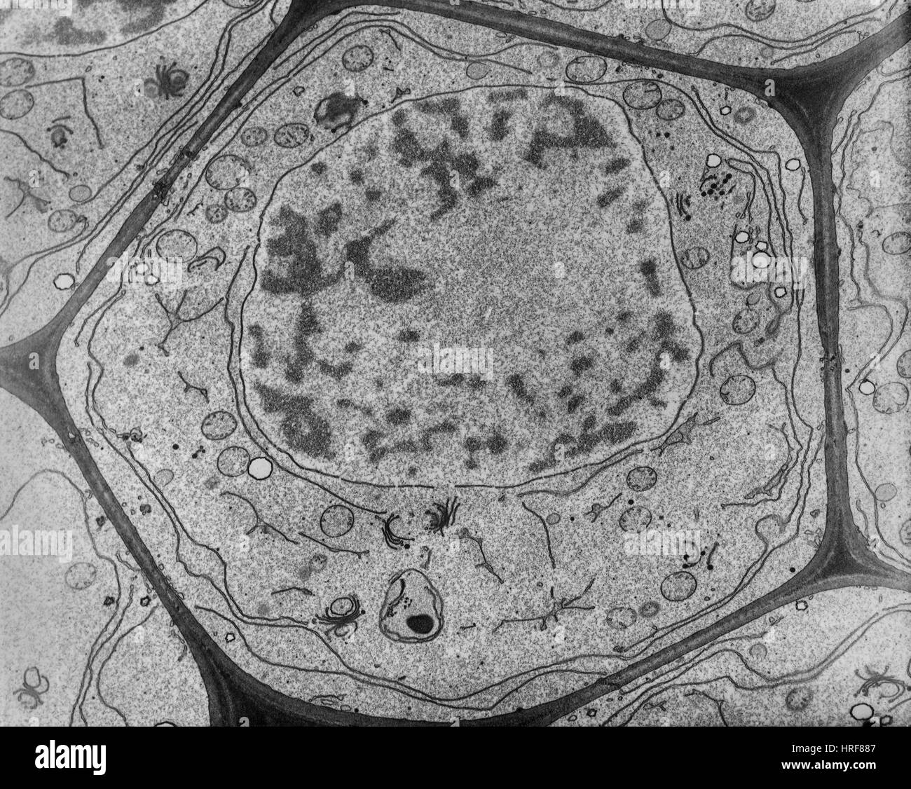
Plant Parenchyma Cell Nucleus Tem Stock Image C036 7371 Science Photo Library from media.sciencephoto.com Both transmission electron microscope (tem) and scanning electron microscope (sem) use electrons to generate images but they differ by the mode of image generation. To study and differentiate between plant and animal cells. See more ideas about electron microscope, microscopic photography, microscopic. Find the perfect transmission electron microscope stock photos and editorial news pictures from getty images. In transmission electron microscope (tem), the source of transmission electron microscopy involves a high voltage beam of electron emitted by a cathode and formed by magnetic lenses. An image of a single cell of the plant pathogenic bacterium, pseudomonas syringae, is presented in fig. The use of a transmission electron microscope (tem) to study minute no colour images: So, if you want to know more about the transmission electron microscope and transmission electron microscopy techniques working principle in detail, stay tuned to this microscopy lecture.
Transmission electron microscope (tem) images.
Light and electron microscopes allow us to see inside cells. Both transmission electron microscope (tem) and scanning electron microscope (sem) use electrons to generate images but they differ by the mode of image generation. This increased resolution allows us to study ultrastucture of organelles, viruses and macromolecules. There are two main types of electron microscopes: A very high resolution for observing and analyzing the composition of elementary structures. Transmission electron microscopy is a proven technique in the field of cell biology and a very useful tool in biomedical research. Using electrons to explore the micro world. Difference between electron beam and light. The transmission electron microscope (tem) operates on many of the same optical principles as the light microscope. Physics of image formation (springer series in optical sciences, vol 36) by l. Visualization of biological molecules in their native state by joachim. Electron microscopes use electron beams focused by electromagnets to magnify and resolve microscopic specimens. On plant leaves, there is a strong selection for cells with traits that would also favour.
Share :
Post a Comment
for "Plant Cell Transmission Electron Microscope Images / Tem Of Plant Cell - Transmission electron microscopy <ul><li>in a conventional transmission electron microscope, a thin specimen is irradiated with an electron."


Post a Comment for "Plant Cell Transmission Electron Microscope Images / Tem Of Plant Cell - Transmission electron microscopy <ul><li>in a conventional transmission electron microscope, a thin specimen is irradiated with an electron."